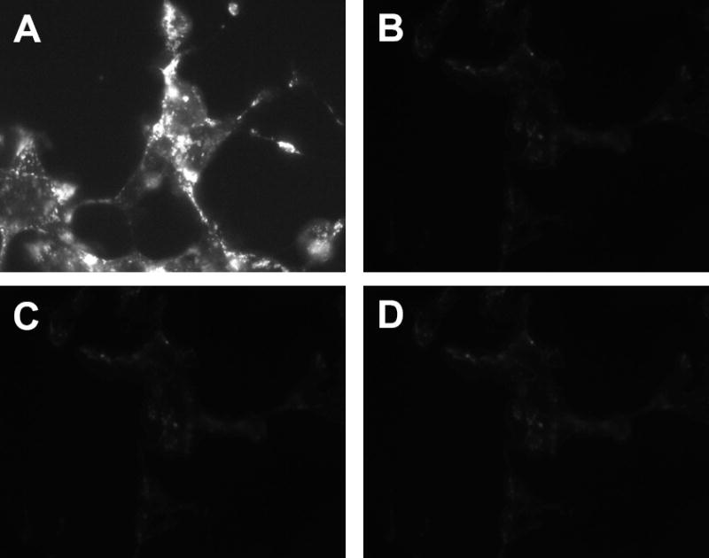
Figure 4. CD63 protein expression is abolished in HEK-293 cells using siRNA.

Panel A: Immunocytochemical localization of endogenous CD63 protein in a G418 selected HEK-293 line that expresses CD63. The experiment was performed at room temperature in presence of saponin. Panel B: CD63 expression in a clonal line of HEK-293 transfected with siRNA targeting CD63 then selected with G418, CD63-knockdown). Panel C: The same as in panel A, but with primary antibody omitted. Panel D: The same as in panel B, but with primary antibody omitted. The anti-CD63 monoclonal antibody (primary antibody) was diluted 1:100. The immunocytochemical localization of CD63 shown in panels A to D was repeated 10 times with similar results.
