
Figure 1.
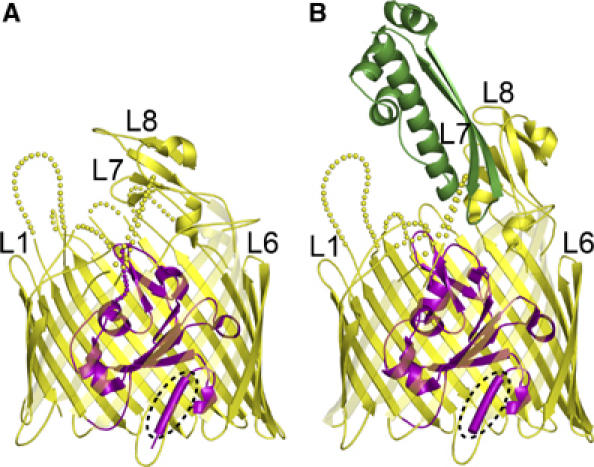
Ribbon diagrams of colicin I receptor (A) and the Cir–colicin Ia complex (B). The extracellular space is located at the top of the figure and the periplasmic space is at the bottom. Cir spans the outer membrane using a 22-stranded beta barrel (yellow) and contains a globular plug domain (magenta) within the barrel lumen. Residues comprising the TonB box (E31–T32–M33–V34–V35) are shown as a tube and encircled by a dotted line. Some extracellular loops are labelled for reference; disordered parts of the loops are indicated by dotted lines. In the Cir–colicin Ia complex, the R-domain of colicin Ia (green) interacts extensively with extracellular loops 7 and 8 of Cir. The fold of the colicin Ia R-domain is identical to that observed in the crystal structure of full-length colicin Ia (Wiener et al, 1997). All figures were prepared with PYMOL (DeLano).