
Figure 5.
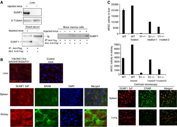
SUMF1 is taken up into mice tissues. (A) Uptake and secretion of SUMF1 in tissues of wild-type mice. AAV2/8TBG-3xFS1 particles (2.8 × 10¹¹ total) were tail-vein injected into three C56BL/6 mice. Ten days later, control (non-injected) and injected mice were killed and their tissues were analyzed. Expression of SUMF1 was detected in the transduced livers by Western blotting and secretion of SUMF1-Flag was detected in the plasma by immunoprecipitation (one transduced mouse shown). The uptake of SUMF1-Flag in bone marrow cells was evaluated by immunoprecipitating cell extracts, and is shown for all of the injected mice. Anti-Flag and anti--tubulin antibodies were used. (B) Immunofluorescence of sections from injected mice. The expression of SUMF1-Flag was analyzed in the transduced liver. Uptake and subcellular localization of SUMF1-Flag were analyzed in the spleen, kidney and lung by fluorescence and confocal microscopy techniques. Anti-Flag polyclonal and anti-ERAB monoclonal antibodies were used. DAPI is shown as a nuclear marker. (C) ARSC activity following uptake of SUMF1 in Sumf1−/− mice. AAV2/8TBG-3xFS1 particles (5.6 × 10¹¹ total) were temporal-vein injected into two Sumf1−/− mice and one wild-type mouse at p2. Ten days later, transduction and ARSC activities were assayed in their liver and kidney homogenates.