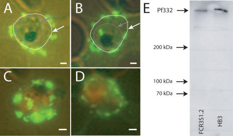
Figure 4. The Pf332 DBL-domain is iRBC surface associated.
Immunofluorescence with antibodies against the DBL-domain visualizes the surface association of the Pf332 DBL-domain as shown for HB3 iRBC (A+C) and FCR3S1.2 iRBC (B+D). A+B: Parasites are unstained; the RBC membrane is indicated with a white line/arrow, the intracellular parasite with a grey line/arrow. C+D: Parasites are stained orange with Ethidium-bromide. Scale bars = 1 µm. E: Western blot with the same α-Pf332 DBL-domain antibody as used for the immunofluorescence assays; the antibody recognizes only the molecule Pf332 and does not cross-react with any other proteins.

