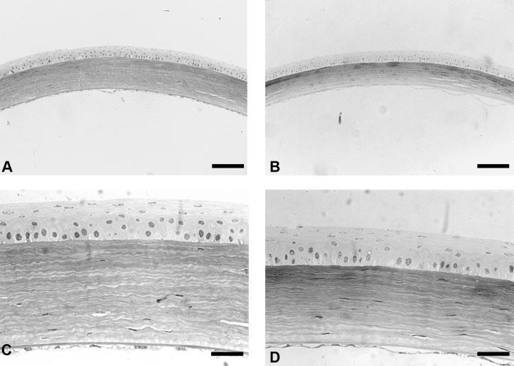
Figure 3.

Light micrographs of wild-type (A, C) and Dpt−/− (B, D) mouse corneas stained with toluidine blue. Dpt−/− corneas were thinner than the wild-type corneas, because of stromal changes (A, B). There was no change in epithelial thickness in Dpt−/−corneas (D) when compared with the wild type (C). Scale bars: 100 μm (A, B) and 33 μm (C, D).
