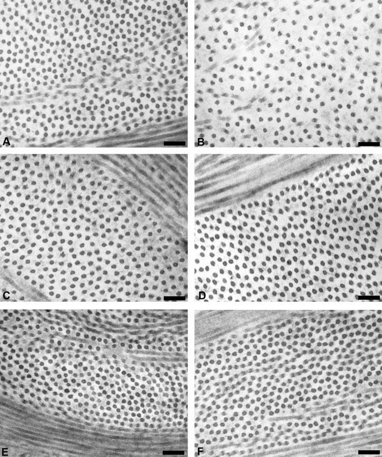
Figure 5.

TEM micrographs of wild-type and Dpt−/− corneas. Collagen fibrils in the posterior stroma of Dpt−/− corneas showed increased interfibrillar spacing (B) compared with the wild-type (A). There was no obvious difference between wild-type and Dpt−/− mid (C, D) and anterior (E, F) stroma. Scale bars, 100 nm.
