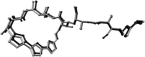
FIGURE 2.
Comparison of rigidly docked structure of kabiramide C into the receptor coordinates of actin in 1YXQ (light shading) against the x-ray coordinates in 1QZ5 (dark shading), after alignment of C-α carbons in actin. RMSD between the two kabiramide structures is 0.71 Å, compared to the C-α RMSD of 0.41 Å.

