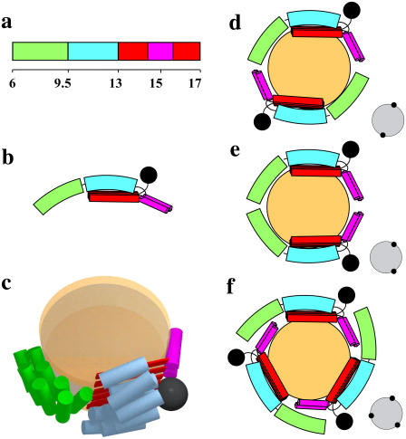
FIGURE 8.
Models of reconstituted B6.4-17/DMPC particles. (a) Domain diagram of B6.4-17. The N-terminal half of the α-helical domain, green; C-terminal half of the α-helical domain, cyan; β-sheets in the C-sheet domain, red; proposed α-helical region in the C-sheet domain missing in the lipovitellin crystal structure, magenta. (b) Proposed domain interactions in lipid-bound B6.4-17. The black sphere indicates a nanogold label. (c) A three-dimensional cartoon of B6.4-17 in a discoidal particle. The orange disks represent a DMPC bilayer. The lipid core and B6.4-17 domains are shown approximately to scale. (d–f) Proposed protein assemblies in the B6.4-17/DMPC particle. (d) Two proteins in a head-to-tail assembly. (e) Two proteins in a head-to-head assembly. (f) Three proteins in a symmetric assembly. The expected STEM views are shown on the right corner of each model.

