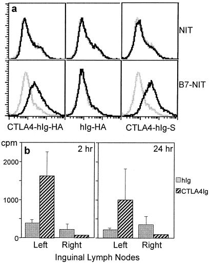
Figure 2.
Fusion proteins bind B7 and accumulate in the draining lymph node. (a) Flow cytometric analysis shows that CTLA4-hIg-HA binds to B7. NIT cells expressing membrane-bound B7-1 or untransfected cells (B7-NIT and NIT, respectively) were incubated with supernatants from Chinese hamster ovary cells transfected with CTLA4-hIg-HA, hIg-HA, or CTLA4-hIg-S (black line). The gray line represents the NIT or B7-NIT cells incubated with untransfected Chinese hamster ovary cell supernatants. FITC-conjugated sheep anti-hIgG was used as the secondary Ab. (b) Enhanced localization of CTLA4-hIg in draining lymph nodes. BALB/c mice were injected in the left quadriceps with radioiodinated CTLA4-hIg or hIg (total cpm was 3 million; total protein was 5 μg). Lymph nodes were harvested at 2 h and 24 h after immunization. The mean and SEM of five mice per group are shown.

