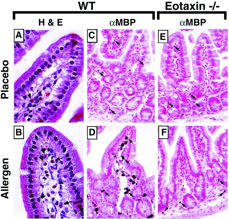
Figure 2.
Histological analysis of jejunum tissue from placebo- and oral allergen-challenged wild-type and eotaxin-deficient mice. (A and B) Photomicrographs represent hemataoxylin- and eosin-stained jejunum sections from placebo- (A) and oral allergen- (B) challenged wild-type mice. (C and D) Photomicrographs represent rabbit anti-mouse MBP [αMBP]-immunostained jejunum sections from placebo- (C) and oral allergen- (D) challenged wild-type mice. (E and F) Photomicrographs represent anti-MBP-stained jejunum sections from placebo- (E) and oral allergen- (F) challenged eotaxin-deficient mice. In placebo-challenged wild-type mice (C), eosinophils are predominantly localized to the crypt region and less frequently within the lamina propria of the villus. In oral allergen-challenged wild-type mice (B and D) but not eotaxin-deficient mice (F), an eosinophilic infiltrate is observed. Eosinophils are present within the mucosa and throughout the length of the villus. Arrows depict representative eosinophils. (A and B, ×950; C–F, ×460.)

