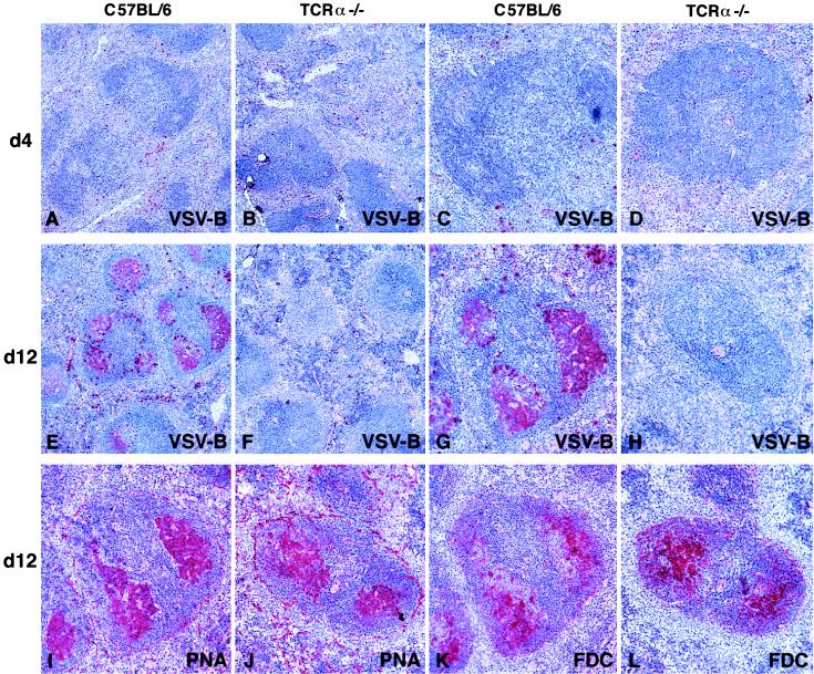
Figure 7.
Lack of VSV-specific germinal center formation in TCRα−/− mice. C57BL/6 mice (A, C, E, G, I, and K) and TCRα−/− mice (B, D, F, H, J, and L) were immunized with 2 × 10⁶ pfu of live VSV i.v. and spleen sections at the indicated time points were analyzed by immunohistochemistry. The stained quality is indicated in each panel: VSV-B, VSV-binding B cells; PNA, peanut hemagglutinin binding; FDC, follicular dendritic cells. Serial adjacent sections of the same follicle are shown from C57BL/6 (G, I, and K) and TCRα−/− (H, J, and L) mice. Original magnifications: ×50 (A, B, E, and F); ×100 (C, D, G, H, I, J, K, and L). Representative results from groups of two to three mice are shown. Similar results were obtained in spleen sections taken on days 20 and 32 after immunization and in three separate experiments.

