Figure 1.
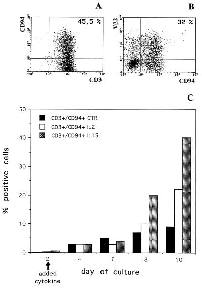
IL-15-induced surface expression of CD94 molecules in TSST-1-stimulated T lymphocytes. PBLs depleted of CD94+ cells were stimulated with TSST-1 (specific for the Vβ2 segment of TCR). IL-15, IL-2, or culture medium was added at 2 days after the initiation of culture. Cells derived from day 10 cultures supplemented with IL-15 were analyzed by double fluorescence for the expression of CD94 and CD3 (A) or CD94 and Vβ2 (B). x axis, green fluorescence; y axis, red fluorescence. (C) Time course of CD94 expression. In this experiments, IL-15, IL-2, or culture medium was added at day 2. Cultures were continued up to day 10, and the proportion of CD3+ cells coexpressing CD94 was evaluated by flow cytometry in cultures supplemented with IL-15, IL-2, or medium, as indicated.

