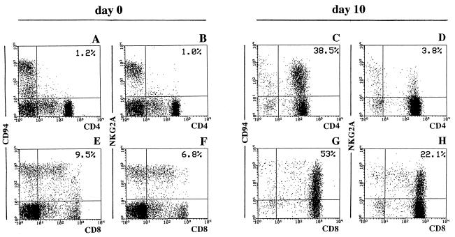
Figure 2.
IL-15-induced expression of CD94 or NKG2A in TSST-1-stimulated lymphocyte populations enriched in CD4+ or CD8+ cells. (A, B, E, and F) Fresh unfractionated peripheral blood lymphocytes were analyzed for the coexpression of CD94 or NKG2A and CD4 or CD8 antigens, respectively. To assess the expression of CD94 or NKG2A in TSST-1-stimulated CD4+ or CD8+ subsets in the presence of IL-15, lymphocytes were depleted of CD4+ or CD8+ cells prior to culture with TSST-1 and addition of IL-15 at day 2. The expression of CD94 or NKG2A by CD4+ (C and D) or CD8+ (G and H) cells cultured for 10 days is shown. Cells have been analyzed by flow cytometry (x axis, green fluorescence; y axis, red fluorescence).

