Figure 1.
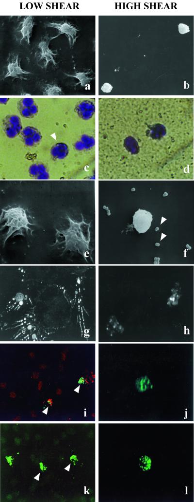
Effects of 60-min exposure to low (0–2 dynes/cm2) and high (>6 dynes/cm2) shear stress on neutrophils adherent to polyetherurethane urea. (a and b) Shear stress causes major morphological changes in neutrophils. Representative scanning electron micrographs demonstrate that adherent neutrophils exposed to low shear were primarily well spread, in contrast to those under high shear levels, which showed condensed and irregular morphologies. (×1,000.) (c and d) May-Grünwald/Giemsa staining revealed that neutrophils under low shear exhibited characteristic multilobed nuclei, with an occasional condensed nucleus (arrowhead in c). Nuclear condensation was observed in all cells exposed to high shear levels. (×100.) (e and f) Shear stress affects the phagocytic ability of adherent neutrophils. Representative scanning electron micrographs show that neutrophils exposed to low shear effectively located and phagocytosed preseeded S. epidermidis. Neutrophils under high shear apparently were incapable of interacting with the bacteria. Arrowheads in f show numerous bacteria in close proximity to a condensed neutrophil under high shear. (g and h) Neutrophil F-actin distribution is influenced by shear stress. F-actin was labeled with rhodamine phalloidin and visualized by using confocal microscopy. (×3,300.) Spread neutrophils in areas of low shear localized actin in pseudopodia, whereas neutrophils exposed to high shear exhibited compact actin distribution and low-intensity staining. (g, ×60, and h, ×3, computerized zoom.) (i and j) Membrane PS is exposed on neutrophils under shear stress. Syto17-counterstained (red) adherent neutrophils exposing membrane PS were identified with annexin V-FITC (green). Areas of low shear contained few annexin V-positive neutrophils, whereas high-shear areas contained sparse but exclusively annexin V-positive neutrophils. (i, ×60, and j, ×3, computerized zoom.) (k and l) DNA fragmentation also was identified after shear stress exposure. By using TUNEL-fluorescein labeling, low numbers of neutrophils with fragmentation were observed under low shear, whereas a high percentage of the sparse cells under high shear exhibited fragmentation (k, ×60, and l, ×3, computerized zoom.)

