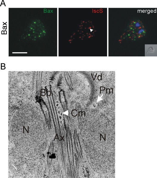
Figure 1. Expression of human Bax in induced Giardia trophozoites does not affect mitosomes.
A) Confocal microscopy analysis of the subcellular distribution of Bax in transgenic cells. Recombinant Bax (left panel, green) does not localize to mitosomes (middle panel, red) labeled with an anti-IscS rabbit antiserum as a mitosome marker. The signature central mitosome structure is indicated with an arrowhead. The merged images show a clear lack of co-localization. Nuclear DNA is stained with DAPI (blue). Inset: differential interference contrast (DIC) image. Scale bar: 5 µm. B) The central mitosome structure (Cm, arrowhead) is resolved as a tightly packed array of spherical organelles in electron microscopy of adherent cells. The subunits are indistinguishable from individual peripheral mitosomes (Pm, arrow). This organelle structure is unchanged in transgenic trophozoites expressing Bax. N, nucleus; Bb, basal bodies; Ax, axonemes; Vd, ventral disk.

