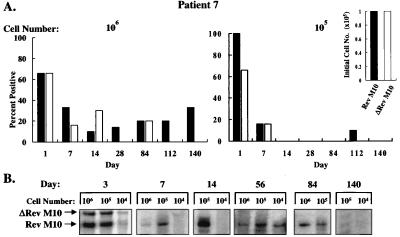
Figure 5.
Detection of pLJ-Rev M10 and pLJ-ΔRev M10 DNA or RNA in patient 7. (A) DNA PCR of 106 (Left) or 105 (Right) cells is shown with relative ratios at the time of infusion presented (Inset). For the Rev M10 vector, 1% of 9 × 10⁹ cells were transduced and for ΔRev M10, 10% of 109 cells were gene-modified. Equal numbers of cells modified with each vector were reinfused (9 × 10⁷). (B) RT-PCR analysis of RNA from PBMCs stimulated with anti-CD3 and anti-CD28 is shown at the indicated times after reinfusion.

