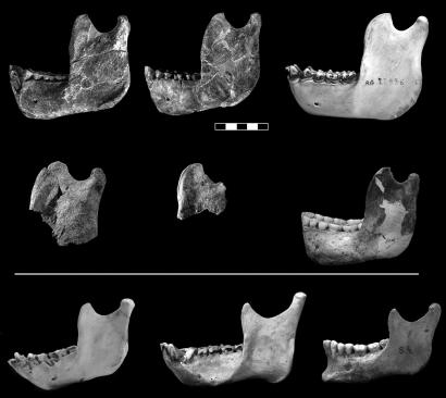
Fig. 1.
Ramal morphology in Au. afarensis and extant primates. (Top) Left mandibular ramus and right mandibular ramus (horizontally flipped) of Au. afarensis specimen A. L. 822-1 and left mandibular ramus of a gorilla. (Middle) Left mandibular ramus of Au. afarensis MAK-VP 1/83 specimen; fragment of left mandibular ramus of Au. afarensis specimen A. L. 333-100; and mandibular ramus of Au. robustus specimen SK 23. (Bottom) Mandibular ramus of a chimpanzee, an orangutan, and H. sapiens. (Scale bar: 5 cm.) Note that the upper end of the ramus in all of the specimens above the white line resembles that of a gorilla (particularly in the shape of the coronoid, the great percentage that the coronoid base constitutes of the ramal width, the confined appearance of the mandibular notch, and the small percentage that the notch area constitutes of the ramal area). The limited reconstruction of the coronoid process on the left ramus of A. L. 822-1 is based on the corresponding preserved area on the right ramus and vice versa.

