Fig. 1.
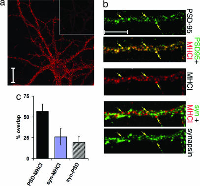
MHCI is expressed at or near synaptic sites. (a) MHCI expression in hippocampal neurons (2 weeks in vitro) detected with Ox18 compared with equimolar amount of control mouse IgG (Inset). Immunoreactivity is present in soma and dendrites. (Scale bar: 20 μm.) (b) High-magnification views of MHCI and synaptic protein immunoreactivity. Antisynapsin antibodies were detected with Cy-2 linked secondary, anti-MHCI with Cy3, and anti-PSD-95 with Cy5. Synaptic proteins are pseudocolored green for comparison with MHCI (red). (Top) PSD-95 immunostained puncta marked by arrows; merged PSD-95 and MHCI signals, with nearly complete colocalization. (Middle) MHCI immunostaining of dendrites and spines. (Bottom) Synapsin immunostaining apposes but does not overlap MHCI immunostaining. (Scale bar: 10 μm.) (c) Quantification of the percentage overlap of MHCI immunostaining (Ox18 antibody) with PSD-95 or synapsin (syn); 57.0 ± 8.4% of MHCI immunoreactive pixels overlap with PSD-95 pixels. However, only 26.1 ± 9.8% MHCI pixels overlap with synapsin pixels. Synapsin–MHCI overlap vs. PSD-95–MHCI overlap is statistically significant (P < 0.001, t test). Overlap between synapsin and PSD-95, 19.3 ± 6.9%. The amount of overlap between synapsin–MHCI vs. synapsin–PSD-95 was not significantly different (P = 0.2, t test) (n = 7 fields of 325 × 325 μm at 512 × 512 pixel resolution).

