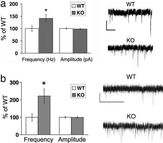
Fig. 2.
Basal synaptic function is altered in β2m/TAP1 KO neurons. (a) Whole-cell recordings from hippocampal neurons in culture. (Left) Frequency of mEPSCs recorded in the basal state from KO (2.5 Hz, n = 17 neurons) is greater than that of WT (1.8 Hz, P < 0.001, n = 22 neurons), but amplitudes do not differ (WT, 7.1; KO, 7.0; P = 0.98). Data presented as ratio of KO/WT median instantaneous frequency (1 per interevent interval) or median amplitude. ∗, significance as calculated by Kolmogorov–Smirnov test of cumulative distribution of events (see Fig. 5 a and b). (Right) Representative traces of mEPSC recordings from WT or KO neurons (2.5 weeks in vitro). (Scale bar: 10 pA; 400 msec.) (b) Whole-cell recordings from layer 4 neurons in visual cortical slices. (Left) mEPSCs from KO are more frequent than WT (WT, n = 10 neurons from three animals; KO, n = 10 neurons from three animals). Frequency (in Hz) (WT, 2.4 ± 1.7; KO, 5.3 ± 3.1; P = 0.02). Amplitude (in pA) (WT, 10.8 ± 1.6; KO, 10.7 ± 1.6; P = 0.92). ∗, significance by t test. (Right) Representative traces of mEPSC recordings from WT or KO neurons. (Scale bar: 10 pA; 400 msec.)

