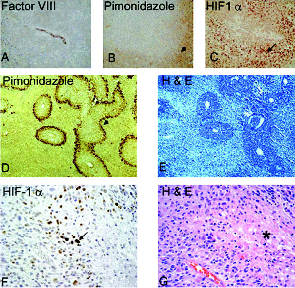
Fig. 1.
HIF stabilization in hypoxic areas. HIF-1α is stabilized in cells distant from a blood vessel. A–C: Adjacent sections of subcutaneously grown tumor xenografts of human LN229 glioma cells. Panel A shows stain Factor VIII, highlighting a vessel. Panel B is pimonidazole staining showing hypoxic areas in brown (arrowhead). Panel C is HIF-1α immunostain (arrow), showing nuclear staining of HIF. D–E: U87 glioma xenografts from a rat orthotopic brain tumor model. Panel D is pimonidazole staining that shows a rim of viable hypoxic cells at the periphery of vascularized regions (arrowhead). Panel E shows the corresponding H & E staining. Note large areas of necrosis (light blue). F–G: Human GBM specimen. Panel F highlights the HIF-1α-positive staining cells localized in the pseudopalisading cells (arrow), and Panel G is the corresponding H & E of the adjacent section showing the necrotic area (asterisk).

