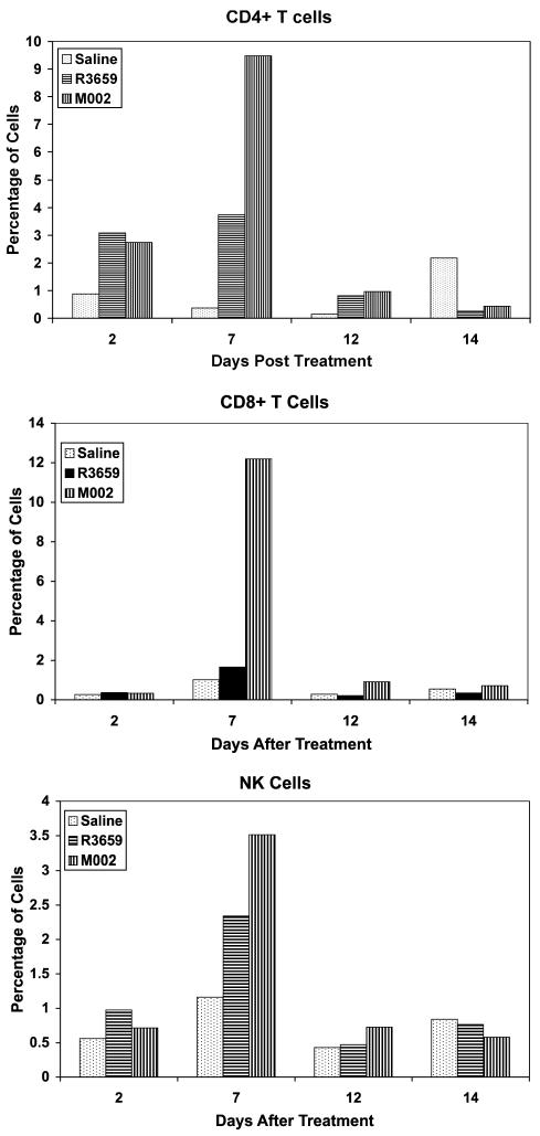
Fig. 5.
CD4+ T cells, CD8+ T cells, and NK cells infiltrating a 4C8 tumor. B6D2F1 mice were induced with 4C8 gliomas that were allowed to grow for 21 days and were then treated intratumorally by injection of 10 μl of either saline, 1 × 10⁷ PFU R3659, or 1 × 10⁷ PFU M002. Two mice from each group were killed at 2, 7, 12, and 14 days after therapy, and the cerebellums were homogenized and filtered through 170-μm pore Nytex (Tetko, Inc., Elmsford, N.Y.) gauze mesh. Aliquots of each were stained with FITC- or PE-conjugated monoclonal antibodies to CD4 T cells, CD8 T cells, or NK cells (GK1.5, 53-6.7, or anti-CD49b, clone DX-5, respectively [Pharmacia]) and subjected to FACS analyses. Data are expressed as percent of gated cells by using side-scatter and forward-angle light scatter indices from similarly treated B6D2F1 spleen cells to define the gate.

