Fig. 3.
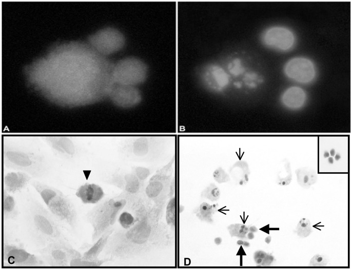
Confirmation of apoptosis induction in glioma cells by a fluorescent microscopy technique and by an in vitro morphologic assay. A. Three CellTracker Green–labeled TALL-104 cells (smaller cells) in contact with a CellTracker Orange–labeled glioma cell (larger cell). B. Hoechst 33342 dye–labeled nuclei of the same cells demonstrating that the glioma cell has a fragmented nucleus. C. H&E-stained brain tumor cell monolayer cultured in the absence of TALL-104 cells demonstrating that the normal, nonapoptotic brain tumor cells are large, contain abundant cytoplasm, and have large oval nuclei. A mitotic figure in a glioma cell at metaphase is seen (arrowhead). D. A view of an H&E-stained brain tumor cell monolayer after coincubation with TALL-104 cells for 4 h. The coincubation of TALL-104 cells with the human brain tumor cells resulted in an increase in apoptotic tumor cells with a concurrent decrease in the number of brain tumor cells with mitotic figures. Cells identified as apoptotic (thin arrows) demonstrated classic morphological changes: either condensed nuclei, fragmented nuclei, or shedding of apoptotic bodies and membrane blebbing. TALL-104 cells were visualized in direct contact with apoptotic brain tumor cells (thick arrows). The inset shows H&E staining of TALL 104 cells.

