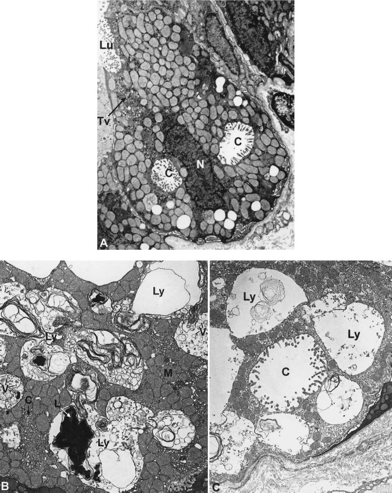
Figure 3.
(A) Electron micrograph of a parietal cell from stomach of a patient with Z-E showing secretory canaliculi (C) and tubulovesicles (TV); Lu, lumen. (Magnification: ×6,500.) (B) Electron micrograph of a parietal cell from the stomach of a patient with ML-IV. The parietal cell lysosomes (Ly) are large and contain structurally heterogeneous material including vesicles (V) and masses of lamellar structures (L). M, mitochondria. (Magnification: ×6,500.) (C) A similar electron micrograph showing an abnormally distended canaliculus that is identified by the cell surface microvilli. (Magnification: ×6,500.)

