Figure 6.
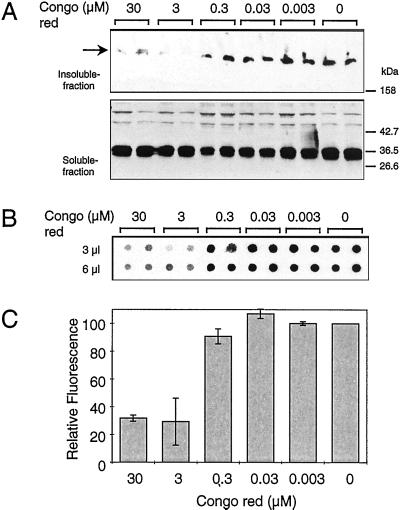
Inhibition of HD exon 1 aggregation in COS cells. (A) Western blot analysis of soluble and insoluble fractions of transfected COS-1 cells after Congo red treatment. COS-1 cells grown for 1 wk in the presence or absence of various concentrations of Congo red (0.003–30 μM) were transfected with the pTL-CAG51 construct. Cell extracts were prepared and fractionated into soluble and insoluble fractions as described in Materials and Methods. Proteins were separated by SDS/PAGE and analyzed by immunoblotting using the HD1 antibody. (B) Filter retardation assay performed on the insoluble fraction of the transfected cell extracts. The SDS-insoluble protein aggregates retained on the filter were detected with the HD1 antibody. (C) Quantitative analysis of the dot blot results shown in B. The dot corresponding to the control experiment without added Congo red was arbitrarily set as 100.

