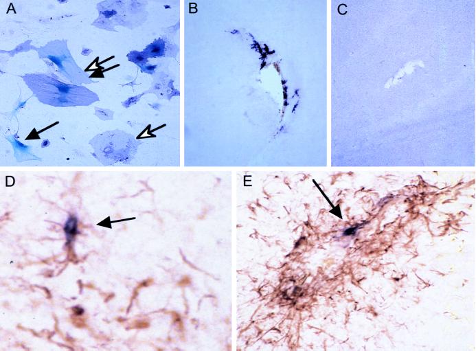
Figure 2.
Gene transfer to transgenic astrocytes. (A) Cultured cells. Primary brain cultures from newborn mice were infected with RCAS-puro, selected for puromycin resistance, then reinfected with a mixture of RCAS-AP and MLV-lacZ (ALV-A) vectors, and stained for AP and β-galactosidase. An open arrow identifies a cell that stains for AP (purple), a solid arrow identifies a cell with nuclear β-galactosidase staining (blue) and the double arrows identifies a cell expressing both markers. (B) Transfer of the AP gene to astrocytes in vivo. Six days after intracranial injection of a newborn Gtv-a transgenic mouse with CEFs producing RCAS-AP, gene transfer can be seen by AP staining in cells with astrocytic morphology around the injection track. (×100) (C) A nontransgenic litter mate injected in the same fashion as in B shows no AP+ cells. (×100) (D and E) GFAP and AP double staining of the injection track 10 days after injection with DF-1 cells producing RCAS-AP demonstrates an increase in number of GFAP+ cells and presence of AP+ cells (arrows). (D, ×100; E, ×200.)

