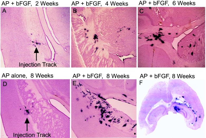
Figure 3.
Time course of proliferation and migration of RCAS-bFGF-infected cells in vivo. An equal mixture of DF-1 cells producing RCAS-AP and DF-1 cells producing RCAS-bFGF was injected into the right frontal lobe of Gtv-a transgenic mice. The mice were sacrificed and brains (40-μm sections) were analyzed for AP activity at 2 weeks (A), 4 weeks (B), and 6 weeks (C) after injection. Mice were injected with DF-1 cells producing either RCAS-AP alone (D) or a mixture of RCAS-bFGF and RCAS-AP (E) and analyzed 8 weeks after injection. (×40.) (F) A representative slice of a brain 8 weeks after infection with RCAS-bFGF and RCAS-AP that shows a diffuse spread of infected cells.

