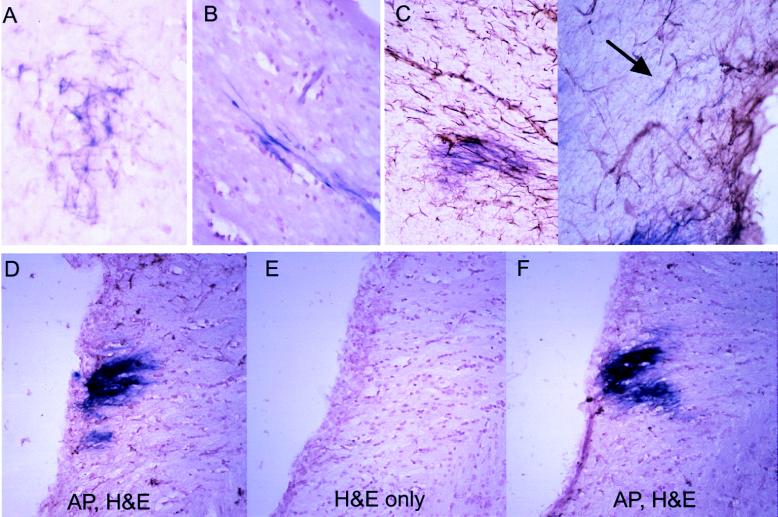
Figure 4.
Morphology and histology of AP+ cells 3 months after infection with RCAS-AP and RCAS-bFGF. (A and B) Astrocytic morphology of infected cells seen in 20-μm sections in gray matter (A) and bipolar morphology in white matter (B) (stained for AP and counterstained with hematoxylin). (C) Staining for AP and GFAP demonstrate a lack of GFAP in most AP+ cells (arrow indicates a rare double-positive cell). (×200.) (D–F) Contiguous hematoxylin/eosin-stained sections with alternate sections also stained for AP. (D and F) AP+ region expected to be found in all three sections. (E) No histologic alteration in the region likely to be AP+. (×100.)

