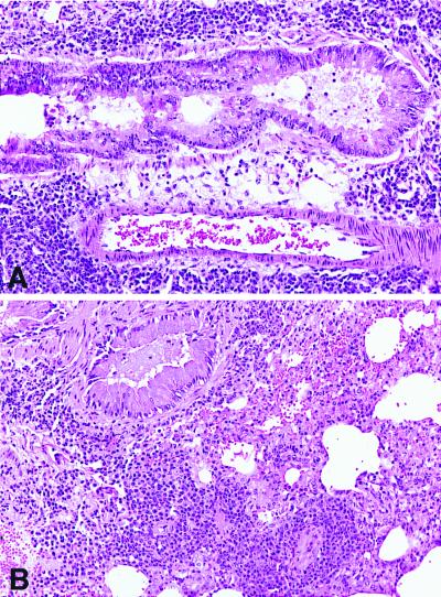
Figure 4.
Histopathology of lung from PDE4D+/+ and PDE4D−/− mice sensitized to OVA. (A) Histology of lung derived from immunized wild-type mice. Sections show dense perivascular and peribronchiolar mixed inflammatory cell infiltrates composed predominantly of lymphocytes and eosinophils. Intraluminal mucous accumulation is noted [hematoxylin and eosin (H&E), ×200]. (B) PDE4D−/− mice. The lungs of PDE4D−/− mice show similar histopathologic changes (H&E, ×200).

