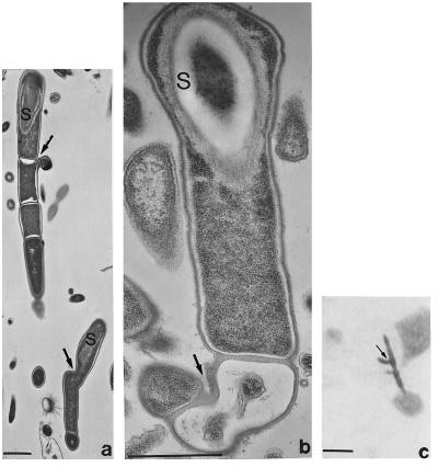
Figure 7.
Branched Arthromitus-like spore formers. (a and b) Transmission electron micrographs; bars = 1 μm. (a) Arrows at branches, third growth region on cell. (b) Early development of the branch point (arrow; S = spore). (c) Branched bacterial filament on unidentified protist; light micrograph from Kirby’s stained slide of Glyptotermes sp. intestine, bar = 5 μm.

