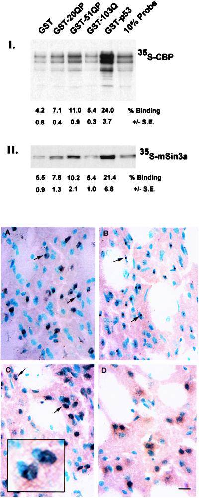
Figure 3.
CBP and mSin3a bind to httex1p in vitro and CBP localizes to nuclear inclusions in R6/2 mouse brains. (Top) Binding of [35S]methionine-labeled CBP or mSin3a to GST-htt fusion proteins. Equivalent amounts of [35S]methionine-labeled CBP (I) or mSin3a (II) were mixed with GST, GST-20QP, GST-51QP, GST-103Q, or GST-p53 attached to glutathione-agarose beads, washed, and the labeled proteins bound to the beads were analyzed by SDS-PAGE. Each experiment was done in triplicate. Percent binding ± SE was calculated by phosphorimager analysis and are listed below each lane. Ten percent of the input of the labeled proteins was also analyzed as shown. (A–D) Localization of CBP to neuronal intranuclear inclusions in R6/2 mice. Striatal sections from R6/2 (A–C) and littermate controls (D) were immunolabeled with anti-htt (A), -ubiquitin (B), and -CBP (C and D) antibodies. Nuclei were counterstained with methyl green. Nuclear inclusions are indicated by arrows. Scale bar, 15 μm.

