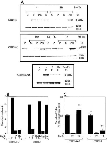
FIG. 4.
(A) Pretreatment of C3H/HeJ peritoneal macrophages with S. enterica serovar Typhimurium products results in tolerization of ERK activation by PG. Macrophages were not pretreated (−) or were pretreated overnight with 107 heat-killed wild-type S. enterica serovar Typhimurium SL1344 bacteria (Hk), 5% (vol/vol) culture supernatant of SL1344 (Sup), 5% (vol/vol) LB broth (LB), 100 ng of LPS per ml (L), or 25 μg of PG per ml (P). They were then left unstimulated (C) or were stimulated for 45 min with 107 live wild-type SL1344 bacteria (S), 25 μg of PG per ml (P), or 1 μg of phorbol myristate acetate per ml (Pm). Cell lysates were prepared and analyzed by Western blotting for the presence of phospho-ERK (p-ERK). Pre-Tx, pretreatment; Tx, treatment. (B) Quantitation of the Western blots shown in panel A. The autoradiographs were scanned and analyzed by using NIH Image software. The mean pixel densities of the phospho-ERK bands (normalized to the pixel densities of the corresponding total ERK bands) are shown. (C) Effect of pretreatment of peritoneal macrophages with heat-killed S. enterica serovar Typhimurium on subsequent TNF-α production in response to PG. Macrophages were not pretreated or were pretreated overnight with 25 μg of PG per ml or with 107 heat-killed wild-type S. enterica serovar Typhimurium SL1344 bacteria. After the pretreatment stimulus was washed away, all groups of macrophages were stimulated for 2 h with 25 μg of PG per ml, and the supernatants were analyzed by ELISA for TNF-α. The normalized amount of TNF-α was expressed as a percentage of the amount produced by the nonpretreated, PG-stimulated cells. The data are the means of the results of three separate experiments, and the error bars indicate standard deviations. The P values for the comparisons are <0.0005 (one asterisk) and <0.0005 (two asterisks).

