Figure 5.
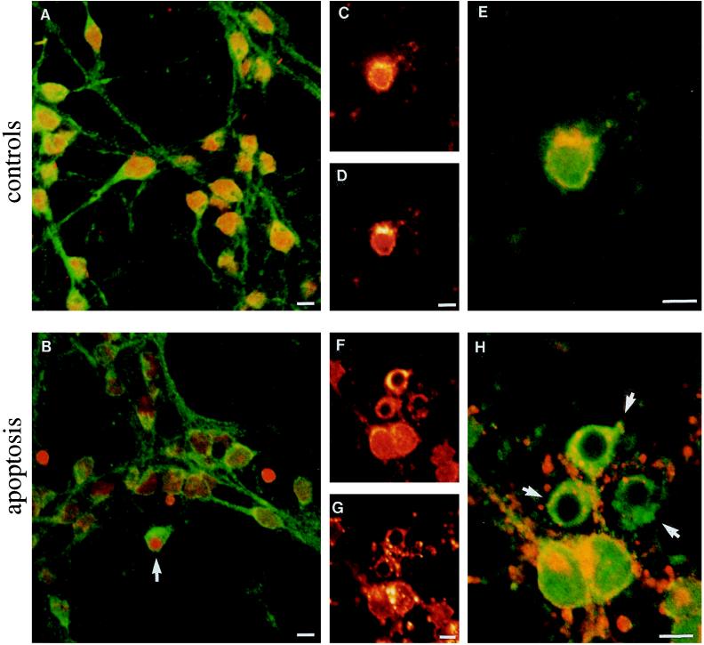
Cytoplasmic condensation of Aβ epitope and fragmentation of Golgi apparatus in cerebellar granule cells undergoing apoptosis. Confocal micrographs of controls (A, C, D, and E) and 6-h apoptotic (B, F, G, and H) neurons are shown. (A and B) Neurons were double-labeled with 4G8 antibody (green) and propidium iodide (staining the nuclei red). (A) In control neurons, 4G8 immunolabeled cytoplasm and neuronal processes. (B) The apoptotic cell (arrow), recognizable by a small strongly stained nucleus, shows 4G8 immunolabeling concentrated in the condensed cytoplasm. Two “naked nuclei” are also seen, whereas the remaining neurons show different degrees of apoptotic modifications. (C, D, F, and G) Cells were double-stained with 4G8 antibody (C and F) and rhodamine-conjugated WGA (D and G). The two chromophores are shown separately with the same color scale. An higher-magnification view of cells shown in C, D, F, and G is, respectively, reported in E and H, where the two chromophores are superimposed in the same image with a green/red color scale. (E) In control neurons, 4G8 (green) and WGA Golgi (red) staining colocalize (yellow), whereas in three apoptotic neurons (H, arrows), the 4G8 signal appears to be mostly condensed in cytoplasm and does not colocalize with the Golgi apparatus, which is fragmented.

