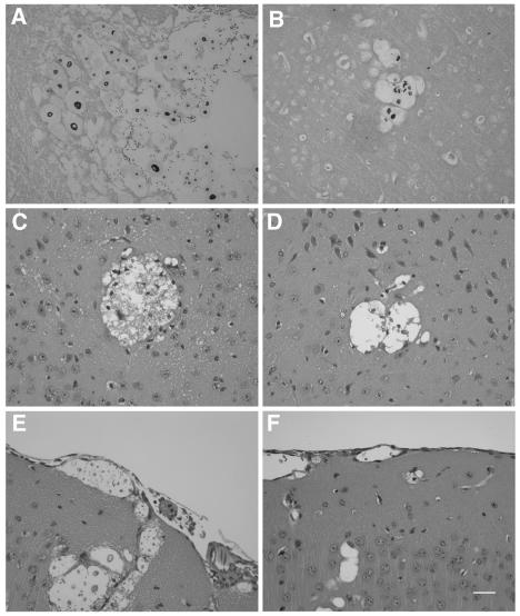
FIG. 5.
Histopathology of mice infected with the wild type (A, C, and E) and the cpra deletion mutant (B, D, and F). Mice were challenged with yeast cells through the tail vein. Brains were removed and fixed in 10% buffered neutral formalin. Tissue sections stained with Gomori methenamine silver (A and B) were from mouse brains 6 days postinjection for panel A and 9 days postinjection for panel B. Tissue sections stained with hematoxylin and eosin (C, D, E, and F) were from mouse brains 9 days postinfection. Neural parenchyma show locular lesions containing yeast cells (A, B, C, and D). The meningeal areas show cystic spaces that contained cryptococci (E and F). Bar, 30 μm.

