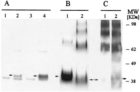
FIG. 1.
Expression of recombinant F2 in E. coli. (A) Recognition of pQE60-Pf-F2 (native codons) and AKI-eF2 (E. coli codons) by a conformational MAb, MAb RII.10, in a Western blot. Lanes 1 and 2, uninduced and induced pQE60-Pf-F2, respectively; lanes 3 and 4, uninduced and induced AKI-eF2, respectively. Monomeric F2 is indicated by the arrows. (B) Coomassie blue-stained polyacrylamide gel comparing the reduced (lane 1) and nonreduced (lane 2) Ni-NTA-purified AKI-eF2, revealing that the protein tends to form aggregates. (C) Western blot analysis revealing that only a small proportion of the monomeric form of AKI-eF2 is recognized by a conformational MAb (lane 1) compared to the polyclonal anti-EBA antibodies (lane 2).

