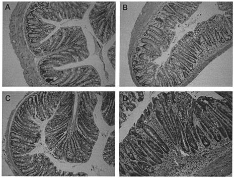
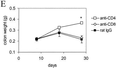
FIG. 4.
Histology and weights of colons from infected mice depleted of CD4+ T cells. The panels (hematoxylin and eosin-stained sections; original magnification, ×200) show representative stained colonic sections from uninfected C57Bl/6 mice treated with rat IgG (A) or CD4-depleting MAb GK1.5 (C) for the previous 4 weeks. Sections from C. rodentium infected mice treated with rat IgG (B) or CD4-depleting MAb GK1.5 (D) for the previous 4 weeks are shown in comparison. There were focal inflammatory cell infiltrates, crypt hyperplasia, disruption of the crypt architecture, and evidence of vasculitis in the submucosa of infected CD4+-T-cell-depleted mice. (E) On day 28 postinfection, the distal colon of infected CD4+-T-cell-depleted mice also weighed significantly more (asterisk, P < 0.05) than the colons of infected CD8+-T-cell-depleted mice or rat IgG-treated control mice.


