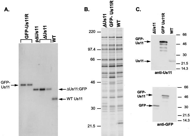
FIG. 7.
Construction and characterization of a recombinant HSV-1 expressing an EGFP Us11 fusion protein. (A) Southern analysis of the Us-TRs junction fragments. Viral (GFP-Us11R, ΔUs11, and WT) or plasmid (pΔUs11) DNA was digested to completion with EcoNI, fractionated by electrophoresis in a 1% agarose gel, and transferred onto a nylon membrane. After the blot was probed with a 32P-labeled NcoI-PflMI DNA fragment derived from the Us-TRs region shown in Fig. 6B, the washed membrane was exposed to X-ray film. The plasmid pΔUs11 was used to construct the recombinant virus ΔUs11. EcoNI-digested viral DNA from two different isolates of ΔUs11 appears as a standard. The genetic structure that surrounds the Us-TRs junction in both the plasmid (pΔUs11) and the ΔUs11 viruses is depicted in Fig. 6C. ΔUs11 was the parental virus from which the GFP-Us11R recombinants were constructed. Two plaque-purified isolates that contain a GFP-Us11 fusion gene as depicted in Fig. 6D are shown. (B) Pattern of late protein synthesis in cells infected with a recombinant virus that expresses a GFP-Us11 fusion protein. Vero cells were infected with WT HSV-1, the ΔUs11 mutant, or the GFP-Us11R virus. At 16.5 h postinfection, the cultures were labeled with 35S-labeled amino acids for 1 h, the samples were solubilized in SDS-PAGE loading buffer, and the proteins were fractionated in SDS-polyacrylamide gels. The fixed, dried gel was subsequently exposed to X-ray film. Molecular mass standards (in kilodaltons) appear to the left of the panel. (C) The GFP-Us11R recombinant virus expresses a GFP-Us11 fusion protein. Total protein was isolated from cells infected with either WT, ΔUs11, or GFP-Us11R HSV-1. After fractionation by SDS-PAGE, the polypeptides were electrophoretically transferred to a membrane and incubated with either anti-Us11 or anti-GFP antibodies. Proteins were detected by chemiluminescence after incubation with horseradish peroxidase-conjugated secondary antibodies. The relative migrations of the GFP-Us11 fusion protein, GFP, and Us11 appear to the left of the image. Molecular mass markers (in kilodaltons) are shown on the right.

