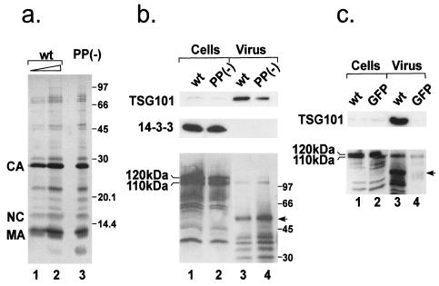
FIG. 8.
Incorporation of cellular proteins into MPMV. (a) wt and PSAP mutant virus was purified by centrifugation through sucrose cushions followed by sedimentation in an Optiprep velocity gradient. wt (lanes 1 and 2) and PSAP mutant (lane 3) virus protein content was analyzed by silver staining. (b) Comparable amounts of lysates from wt and mutant virus-expressing cells (lanes 1 and 2) as well as highly purified virus (the same amounts as in panel a, lanes 1 and 3) were probed for the cellular proteins TSG101 (upper panel), 14-3-3γ (middle panel), and Nedd-4 (lower panel) by Western blotting. In the lower panel, the major Nedd-4-related bands migrating at 120 and 110 kDa are indicated. The arrow marks a band reacting with Nedd-4 antiserum which does not comigrate with one of the major viral proteins detected in the Ponceau S stain (not shown). (c) To analyze whether TSG101 and Nedd-4 are released from 293T cells in the absence of MPMV expression, we probed sucrose pellets from culture media of cells transfected with pMPMV or EGFP expression vectors (lanes 3 and 4) for TSG101 (upper panel) and Nedd-4 (lower panel). The arrow marks the same product as in panel b.

