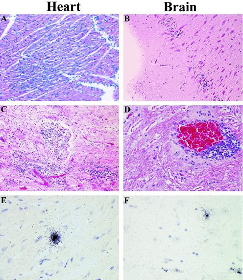
FIG. 4.
EMCV-induced pathological changes and detection of viral RNA in pigs. Five-week-old pigs were intraperitoneally inoculated with 2.9 × 10⁸ PFU of EMCV 30, and histopathologic changes and the presence of EMCV RNA in the heart, brain, spleen, pancreas, liver, kidney, and skeletal muscle were analyzed 7, 21, 45, and 90 days p.i. In the acute phase (7 days p.i.), inflammation and degenerative changes were observed in the heart (A), and brain (B), but no changes were observed in the other organs. In the chronic phase (21 to 90 days p.i), infiltration of lymphocytes in the heart (C) and perivascular cuffing in the brain (D) were evident throughout the infection period up to day 90 p.i. and were accompanied by persistence, as demonstrated by localization of EMCV RNA by in situ hybridization (E and F). For histopathology, heart and brain sections were embedded in paraffin, and 4-μm-thick sections were stained with hematoxylin and eosin. In situ hybridization was performed by using a 35S-labeled VP1 cDNA probe.

