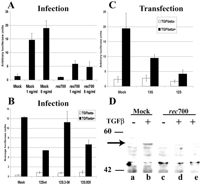
FIG. 5.
E1A inhibits TGF-β-induced signal transduction in adenovirus-infected cells as determined by using the 3TP-lux reporter plasmid. (A) HepG2 cells were transfected with 3TP-lux and pCMV-βGal plasmids. After 6 h of transfection, cells were infected with rec700 at 50 PFU/cell or mock infected. Infections were maintained in the presence of AraC. Cells were treated with TGF-β1 from 12 to 26 h p.i.; subsequently, cells were lysed and luciferase and β-Gal activities were measured. Luciferase values were normalized against β-Gal activity for each sample. Each experimental condition was done in triplicate, and the average values are shown. (B) HepG2 cells were transfected with 3TP-lux and CMV β-Gal plasmids and an empty vector or a plasmid expressing either the 13S or 12S isoform of E1A. At 18 h posttransfection, cells were treated with recombinant TGF-β1 (3 ng/ml) for 8 h. (C) HepG2 cells were treated as described for panel A. Cells were maintained from 12 to 48 h p.i. in the presence of 5 ng of TGF-β1/ml. (D) A549 cells were infected with 50 PFU/cell of rec700 or mock infected and maintained in the presence of AraC. At 20 (lane d) or 24 (lane e) h p.i. cells were mock treated or treated with 5 ng of TGF-β1/ml for 20 min. Levels of phospho-Smad 2 (arrow) were determined by Western analysis. Molecular weight marker positions are shown.

