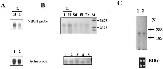
FIG. 3.
Expression and tissue distribution of VIRP1 mRNA in tomato. mRNA extracted from 100 μg of total RNA was size separated in 1% agarose-formaldehyde (A and C) or agarose-guanidine thiocyanate (B) gels. RNA was blotted and hybridized with an α-32P-labeled 0.7-kb VIRP1 antisense RNA probe. (A) Northern blot of RNAs from healthy (lane H) and PSTVd-infected (lane I) tomato leaves. The same blot was also probed with an actin RNA transcript for loading controls as shown in the lower panel (lanes 1 and 2, respectively). (B) Northern blots of RNAs from various tissues. Poly(A)+ mRNA from leaf (L, lanes I [infected] and H [healthy]), seed (lane Sd), flower (lane Fl), and fruit (Fr) were hybridized with antisense VIRP1 RNA. M, DNA molecular weight marker. (C) Northern blot of total RNA from petals (lane 1) and sepals (lane 2) of infected plants hybridized with VIRP1 DNA. Equal loading was verified by visualization of 28S and 18S rRNAs on the blot.

