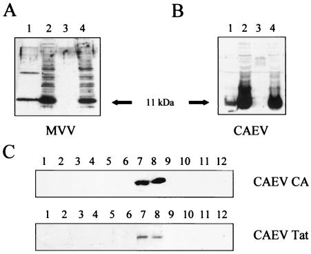
FIG. 3.
MVV and CAEV Tat proteins are associated with viral particles. TIGEF were inoculated (MOI, 1) with MVV (A) or CAEV (B and C) or were mock infected. At 24 h postinoculation, the cells were transfected with pTat-Flag-MVV (A) and pTat-Flag-CO (B and C) plasmids. At 48 h posttransfection, the supernatants were collected and virions were harvested from pellets by ultracentrifugation; cell lysates were then obtained following treatment with the lysing solution. (A and B) Proteins from the supernatants (lanes 1 and 3) and cell lysates (lanes 2 and 4) were analyzed by immunoblot assay by using an antibody directed against the FLAG epitope (diluted 10,000 times). Lanes 1 and 2, cells infected with MVV (A) or CAEV (B); lanes 3 and 4, mock-infected cells. (C) Pellets of virions were resuspended and loaded on the top of a linear sucrose gradient. After 16 h of ultracentrifugation, 12 fractions were collected and proteins were extracted and then analyzed by immunoblot assay with an anti-p25 Gag antibody to reveal the major CAEV capsid protein (diluted 1,000 times) or an anti-FLAG antibody (diluted 10,000 times) to reveal the CAEV Tat protein.

