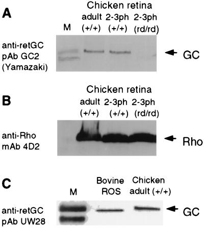
Figure 2.
Analyses of GC1 protein levels expression in normal and rd/rd retina. (A) Western blot probed with anti-retGC pAb GC2. (B) Western blot shown in A reprobed with anti-rhodopsin mAb 4D2. (C) Western blot probed with anti-retGC pAb UW28. The relative mobility of the chicken GC1 polypeptide is slightly slower than that of the bovine GC1 polypeptide. Lane M in A and C shows molecular mass markers, 115 and 80 kDa. 2–3ph, 2–3 days posthatch; ROS, rod outer segments.

