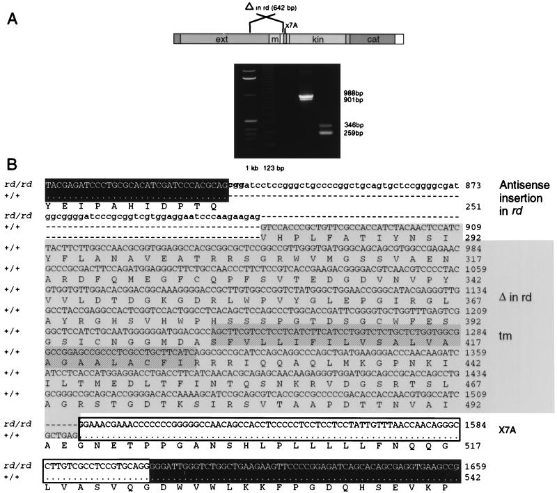
Figure 5.
Identification and analyses of the rd mutation. (A) RT-PCR analyses of +/+ and rd/rd total retinal RNA. The larger fragment in both the +/+ and rd/rd samples contains the alternatively spliced 87-bp nucleotide sequence. (B) Comparison of the +/+ and rd/rd GC1 cDNA sequences at the site of the rd mutation. Sequences shown in white letters on black are identical in +/+ and rd/rd. The 81-bp insert found in the rd/rd GC1 cDNAs is shown in lowercase letters. In some rd/rd GC1 clones, the 3 bases at the 5′ end of the fragment (CGG, bold text) were not present. The sequence corresponding to putative exons 4–7 in +/+, which is deleted in rd/rd, is shaded. The putative transmembrane domain is hatched. The boxed sequence, corresponding to putative exon 7A, is alternatively spliced in both rd/rd and +/+ GC1 transcripts. Residues identical in rd/rd and +/+ GC1 sequences are shown as dots; residues missing from either sequence are shown as hyphens.

