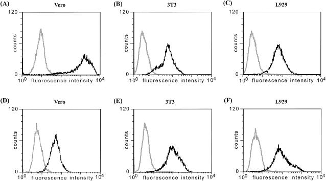
FIG. 3.
Surface expression of PRV gC and gD on ORFV recombinant-infected cells. Flow cytometry of the indicated cells 24 h after infection with D1701-VrVgC (A to C) or D1701-VrVgD (D to F). Nonfixed infected cells (dark lines) or noninfected cells (bright lines) were stained 24 h after infection with the antigen-specific antibodies. The diagrams show the number of counted cells exhibiting specific fluorescence intensities.

