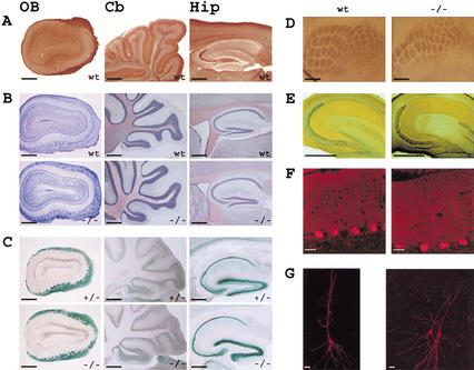
Figure 2.
Brain anatomy in STOP−/−, STOP+/−, and wild-type adult mice. (A) STOP distribution in the olfactory bulb (OB), cerebellum (Cb), and hippocampus (Hip) from wild-type mice (wt). Parasagittal brain sections were stained with 23C STOP antibody. (B) Cell layer organization in wild-type (wt) and STOP−/− (−/−) mice, in the brain regions shown in A. Parasagittal brain sections were stained with cresyl-violet. (C) LacZ expression in parasagittal brain sections from heterozygous mice (+/−) and STOP−/− (−/−) mice, revealed by β-galactosidase activity. (D) Barrel field organization of the somatosensory cortex in wild-type (wt) and STOP−/− (−/−) mice. Tangential brain sections were stained to reveal the cytochrome oxidase activity pattern. (E) Mossy fiber organization in wild-type (wt) and STOP−/− (−/−) mice. Staining for zinc by the Timm sulfide-silver method showed similar mossy fiber pathway in wild-type and STOP−/− mice. (F) Dendritic organization of Purkinje cells in wild-type (wt) and STOP−/− (−/−) mice. Cerebellum sections stained for calbindin indicated normal dendritic arborization in STOP−/− mice. (G) Representative examples of CA1 pyramidal cells in wild-type (wt) and STOP−/− (−/−) mice. The pictures were obtained by confocal imaging of neurons intracellularly labeled with neurobiotin. The pyramidal cells shown in STOP−/− were both injected. A total number of 27 STOP−/− CA1 pyramidal cells were examined and exhibit normal somatodendritic organization. Bars, A–E, 0.5 mm; F, 20 μm; G, 40 μm.

