
Figure 4.
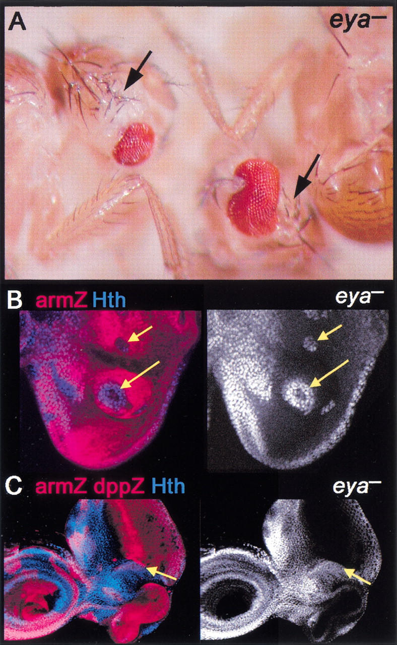
eya represses hth and head capsule development. (A) Adult flies containing eya− clones. Although eya− tissue is unmarked, these heads show a loss of eye and a corresponding increase in head capsule (arrows). (B,C) eya− clones, marked by the absence of arm–lacZ (red), stained for Hth (blue in left panels and white in right panels). Hth is de-repressed cell-autonomously in eya− cells. In C, the disc also expresses dpp–lacZ (strong red stripe). Hth is de-repressed in eya− cells, even when they are adjacent to Dpp-expressing cells (yellow arrows in C).