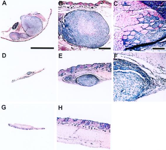
Figure 2.
Effect of PV1(RIPO) on s.c. HTB-15 tumor xenografts in athymic mice. Mice received bilateral implants. When tumors reached an average cross diameter of 8 mm (6–8 weeks after implantation), the mice were given a single i.v. injection of PV1(RIPO) (n = 10) or PBS (n = 10). An s.c. tumor of a PBS-control-treated animal 2 weeks after PBS treatment (A) infiltrated into surrounding tissues (B) and skeletal muscle (C). The tumors regressed 2 weeks after i.v. administration of 2 × 10⁷ pfu PV1(RIPO) (D), revealing remaining tumor mass encircled by necrotic debris and inflammatory infiltrates invading the tumor from the periphery (E and F). At 3 weeks after PV1(RIPO) treatment, the tumors had been replaced by a fibrotic patch (G) with no evidence of residual neoplastic cells (H). Sections are 12 μm thick; hematoxylin/eosin stain. [A, Bar = 8 mm (applies to A, D, and G); B, Bar = 1 mm (applies to B, E, and H); C, Bar = 0.2 mm (applies to C and F)].

