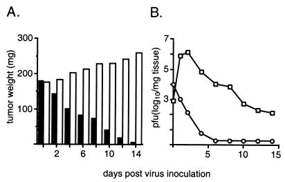
Figure 3.
Intratumoral propagation of PV1(RIPO) in s.c. HTB-15 xenografts grown in athymic mice. (A) Tumor growth in mock-treated (open bars) animals and xenograft regress in mice treated with PV1(RIPO) (closed bars). Mice (n = 4) with similarly sized tumors (8-mm cross diameter) were treated either with a single i.v. inoculation of 2 × 10⁷ pfu of PV1(RIPO) or with PBS alone and were killed at the indicated intervals. Tumors were dissected, weighed, and processed for determination of the viral load by plaque assay (10). The data shown represent mean values for all four animals comprising an experimental group. At 2 weeks after virus treatment, tumor tissue could no longer be macroscopically discerned (compare Fig. 2). (B) Intratumoral (squares) and i.v. (circles) virus load after i.v. administration of PV1(RIPO) to xenografted athymic mice.

