Figure 4.
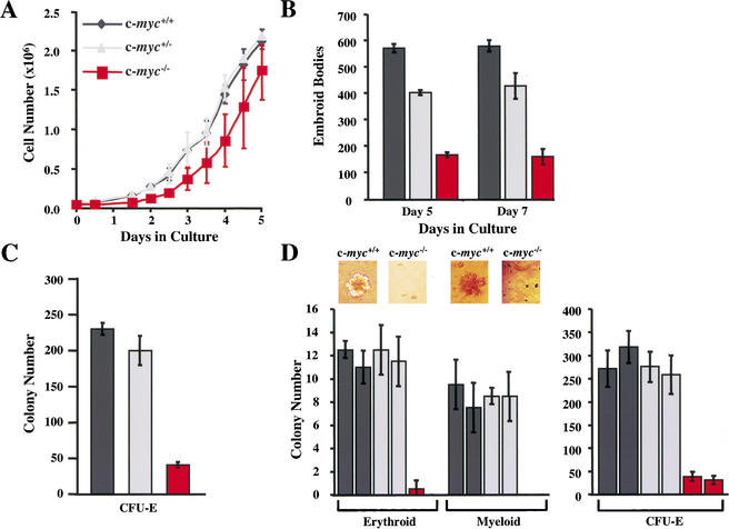
c-myc−/− ES cells are impaired in their in vitro differentiation. (A) Growth rates of wild-type (dark gray), c-myc+/− (light gray), and c-myc−/− (red) ES cells are shown in triplicate. (B) c-Myc deficiency impairs the primary differentiation of ES cells. The indicated ES cells were induced to undergo primary differentiation and after 5 or 7 d in culture, the numbers of EBs were determined. The results shown are the mean of three experiments performed in duplicate. (C) c-myc−/− ES cells are selectively impaired in secondary CFU-E colony formation. The indicated cells, wild-type (dark gray), c-myc+/− (light gray), and c-myc−/− (red), were plated in methylcellulose for 7 d. Then the EBs were dispersed and plated in methylcellulose containing Epo (CFU-E). Colony numbers were determined after 4 d. The data shown are representative of three experiments performed in duplicate. The mean number of colonies +/− the standard deviation is shown. (D) Hematopoietic progenitor colony assays were performed on E9.5 wild-type and c-myc−/− yolk sacs cultured in methylcellulose. Examples of erythroid-like and myeloid-like colonies formed by wild-type and c-myc−/− yolk sac cells in the absence of cytokines are shown above the left panel. In the right panel is shown CFU-E colony assays performed on individual E9.5 yolk sacs isolated from wild-type, c-myc+/−, and c-myc−/− yolk sacs, performed in duplicate.

