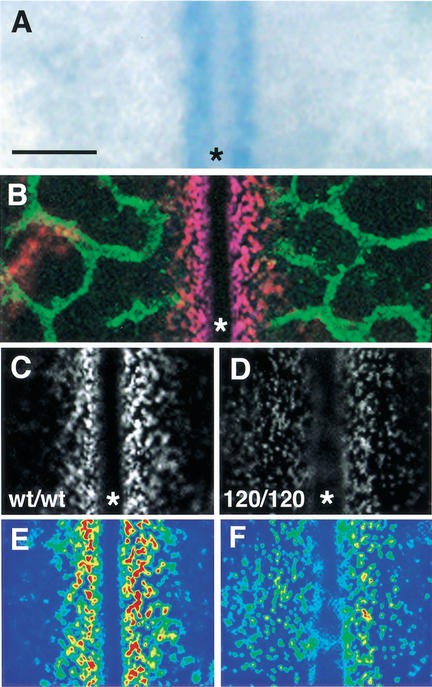
Figure 3.
Altered distribution of secreted VEGF-A protein in the absence of heparin-binding isoforms. (A) VEGF-A gene expression near the hindbrain midline at 10.5 dpc, monitored with a VEGF-A lacZ reporter. (B) At 10.5 dpc, extracellular VEGF-A protein (red) concentrates near the midline, and the PECAM-positive microvessel network (green) is closing in on the VEGF-positive area. (C,D) Comparison of VEGF-A protein distribution in the midline region of wt/wt (C) and 120/120 (D) hindbrains at 10.5 dpc. (E,F) Pseudo-coloring of C,D to highlight regions of highest (red) and lowest (blue) staining intensity. The midline is labeled with an asterisk. Bar, 50 μm.

